International Journal of Arrhythmia 2013;14(4): 34-39.
Untitled Document
ECG & EP CASES
Ventricular Tachycardia Ablation in a Patient With Arrhythmogenic Right Ventricular Cardiomyopathy
Kyoung-Min Park, MD, PhD Division of Cardiology, Department of Internal Medicine, Konkuk University Hospital, Konkuk University School of Medicine, Seoul, Korea
Introduction
Arrhythmogenic right ventricular cardiomyopathy
(ARVC) is an inherited myocardial disease
characterized by replacement of the myocardium
by fibrous and fatty tissue that predisposes one
to ventricular arrhythmias and sudden cardiac
death. Ventricular tachycardia (VT), occurring in
up to 64% of ARVC patients usually originates in the right ventricle (RV) and exhibits left bundle
branch block morphology.1 Reentry is the predominant
mechanism, as suggested by the fact
that tachycardias can be initiated by programmed
stimulation and that they can be entrained in
patients with stable VT. In this case, substrate
mapping and pace-mapping were used to identify
reentry circuits because the patient’s vital signs
were unstable during clinical VT.2
Case Report
We report the case of a 65-year-old man with
a history of implantable cardioverter-defibrillator implantation (ICD) due to several episodes of sustained
VT with syncope that required termination
by cardioversion. Two-dimensional echocardiography
and ventriculography revealed diffuse and
severe RV enlargement, slightly reduced RV systolic
function, and normal left ventricular dimension
and function. Cardiac magnetic resonance
imaging (MRI) showed a dilated RV with mild RV
dysfunction and DE of the anterior RV free wall.
These findings were consistent with a diagnosis
of ARVC based on published criteria. The patient
did well on sotalol with good functional status.
However, he began to have palpitations with a
syncopal history. Recurrent VTs were documented,
which were terminated by pacing several
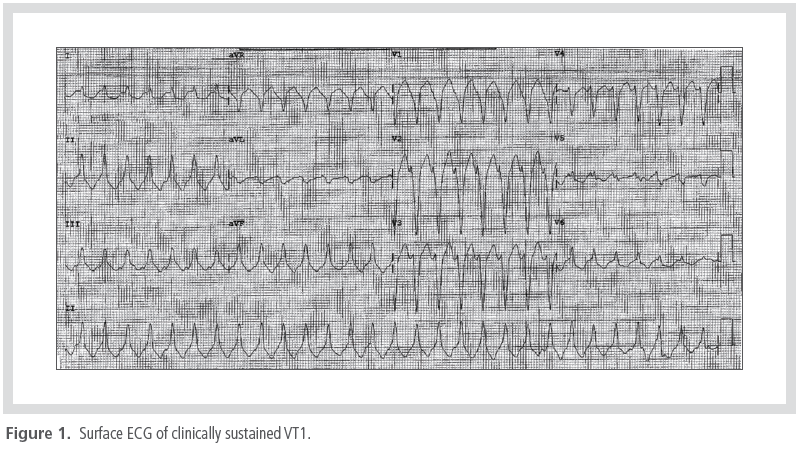
times and by shock on one occasion. The clinical
VT showed the same morphology: left bundle
left-inferior (LBLI) axis, V5 transition, and a 330
msec cycle length with presyncopal attack during
VT (Figure 1). The baseline heart rhythm was
sinus rhythm. During sinus rhythm, the 12-lead
ECG exhibited a localized prolongation (110 msec)
of the QRS complex and inverted T waves with epsilon waves in leads V1 and V2.
First, electroanatomic mapping of the RV was
performed using a 7-French, 4-mm tip ablation
catheter (CARTO® 3, Biosense Webster,
Inc., Diamond Bar, USA) during sinus rhythm in
order to identify the VT substrate on the voltage
map. The voltage map revealed a low voltage
area (<1.5 mV) in the free wall of the RV outflow
tract, peritricuspid area, and apex. Next, a
clinical sustained VT1 with a left bundle branch
block and superior axis QRS morphology (cycle
length = 340 msec) was induced by double extrastimuli
from the RV outflow tract. During VT1,
vital signs were unstable, and therefore, endocardial
bipolar voltage mapping was performed
during sinus rhythm. Endocardial voltage mapping
in the RV revealed anterolateral wall scarring
near the tricuspid annulus (TA) (Figure 2). A
good pace-map was identified near the annulus
at the superior border of the scar. Slightly lower
down, there was a long stimulus-to-QRS with
the same good pace-map morphology (Figure 3).
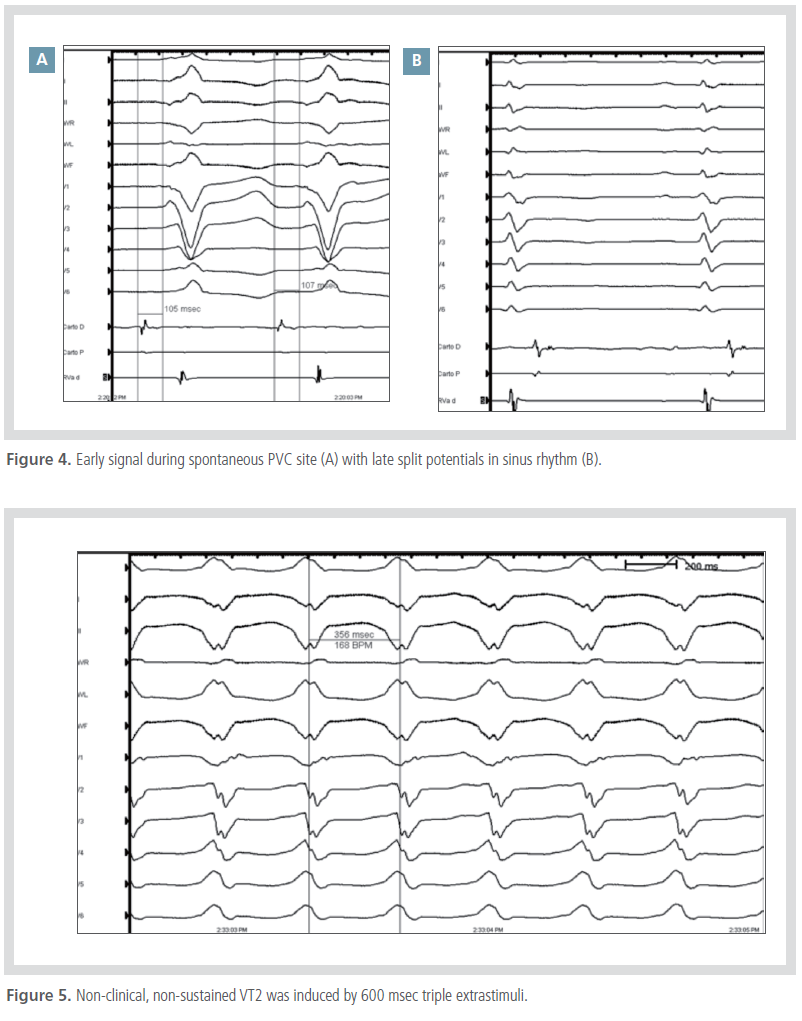
There were early signals at the distal tip of the
ablation catheter, over 100 msec, during clinical
premature ventricular contraction (PVC) at the
site of previous pace-mapping (Figure 4A). The
activation map, at this earliest site, revealed isolated
potentials, which were separated from the
ventricular EGM by an isoelectric line (Figure 4B).
During the clinical VT1 induction study, a nonclinical,
non-sustained VT2 (cycle length = 340 msec) was induced by 600 msec triple extrastimuli
with a 250 msec cycle length (Figure 5). The
excellent pace-map for VT2 was slightly lower
down on the annulus than that of VT1. Our ablation
strategy was to make several lines between
the scar borders to the TA annulus linearly with a
transverse line from the lateral scar border to the
anterior (Figure 6).
Radiofrequency (RF) applications of 60 s each with a target temperature of 50°C and maximum
power output of 50 W were delivered around these
sites. After RF ablation, 400 triple extrastimuli
on 3 ㎍/kg/min isoproterenol only obtained the
non-sustained VT2 whereas the clinical VT1 was
non-inducible. Excellent pace-maps for VT2
were identified on the annulus slightly lower down
from the site of the previous perfect pace-map of
clinical VT1. Several RF ablations were delivered around these good pace-map sites. Eventually,
the VTs could no longer be induced by any pacing
maneuver. No additional epicardial procedure was
performed since both the clinical and non-clinical
VTs were not inducible after the successful endocardial
RF ablations.
Discussion
Tachycardias in the setting of ARVC are inducible
with programmed stimulation and can be
entrained. Reentry involving regions of abnormal
EGMs is the most likely mechanism.
Therefore, the same mapping principles discussed
for idiopathic dilated cardiomyopathy
(IDCM) can be applied in patients with ARVC.
Most VT sites of origin cluster within the lowvoltage
peritricuspid and/or peripulmonic region,
usually within 2~3 cm of the valve’s orifice.3 In
patients with larger scars, the VT can exit toward
the apical extent of the scar, but still within
the region of abnormal EGM voltage. Therefore,
these are the regions initially targeted by activation,
entrainment, and pace-mapping. Activation
and entrainment mapping can be used in hemodynamically
stable VT.
Presystolic activity at the earliest activated
site usually precedes the QRS by at least 30~50
msec.3, 4 Its participation in the reentry circuit
should be confirmed by entrainment. In patients
with noninducible or untolerated VTs, substrate
mapping and pace-mapping can be used to delineate
the region of low-voltage electrogram and then to identify putative components of the
reentry circuits. The acute success rate of ablation
ranges between 50% and 90% in different studies.
The variable reported outcomes can be attributed
to differences in mapping techniques, endpoints,
and operator experience. Overall, substratebased
approaches are associated with better acute
and long-term success rates, a finding that has
been attributed to the patchy distribution of the
scar, harboring multiple regions of slow conduction.
In cases where there are more extensive epicardial
than endocardial substrates, a more aggressive
ablation approach targeting both the
epicardium and endocardium, is often required.
This study clearly indicates that combined endocardial
and epicardial ablation is associated with
better long-term results in terms of freedom from
arrhythmia recurrence.
References
Muir AR, Elliott PM. Arrhythmogenic right ventricular cardiomyopathy.
In: Saksena S, Camm AJ, editors. Electrophysiological disorders
of the heart. 2nd edn. Philadelphia: Elsevier Saunders, 2012;845-853.
Issa ZF, Miller JM, Zipes DP. Ventricular tachycardia in arrhythmogenic
right ventricular cardiomyopathy-dysplasia. In: Clinical arrhythmology
and electrophysiology: A companion to Braunwald’s heart disease.
2nd edn. Philadelphia, PA: Elsevier Saunders, 2012;625-639.