Introduction
Radiofrequency catheter ablation (RFCA) is an established and effective treatment method for various atrial tachyarrhythmias. Although a thromboembolism has been widely reported as the principle complication of conventional RFCA, bleeding complications, including intramural hematomas, have been less often reported [1]. Intramural atrial hematomas are rare, but they have been described as occurring both spontaneously [2] and iatrogenically [3,4]. Here we report the case of a patient with an intramural hematoma associated with RFCA for intractable atrial tachyarrhythmias, who was treated with open cardiac surgery.
Case
A 70-year-old man with a 3-year history of medically intractable paroxysmal atrial tachycardia (AT) and paroxysmal atrial fibrillation (AF) with symptoms of severe palpitation and near-syncope was admitted to the Arrhythmia Center of Seoul National University Hospital.
He had hypertension and was taking an angiotensin-receptor blocker, a beta-blocker, amiodarone, and warfarin. His palpitation continued in spite of treatment with various antiarrhythmic agents, and amiodarone-associated hypothyroidism was suspected. Several months previously, his symptoms worsened compared to those experienced earlier. He visited the emergency department twice within a 4-month period, and cardioversion was performed at every visit. Figures 1A-1D show his various types of atrial tachyarrhythmias.
An electrophysiological study revealed that the atrial tachycardias originated from six different focal sites. Catheter ablation was performed with an electroanatomical mapping system (CARTO-3, Biosense) at the area of the vein of Marshall, left atrial (LA) septum, and cavotricuspid isthmus. Circumferential antral ablation was performed on four pulmonary veins, including the carina, for AF ablation. After RFCA for AF and AT, three types of focal ATs were sustained and continuously changing from one type to another. Thus, DC cardioversion (5 J, internal) was performed to convert to sinus rhythm.
After the first RFCA, he visited the emergency department four times within a 5-month period, and cardioversion was performed at three visits. Five months later, he was admitted for a second RFCA procedure. He arrived at the EP laboratory with a sinus rhythm. Systemic anticoagulation with intravenous heparin was initiated immediately after transseptal puncture. Activated clotting time (ACT) was checked every 30 minute to ensure it remained at 300-350 s.
Three different ATs were easily induced with programmed electrical stimulation. A three dimensional activation map was prepared using the CARTO-3 (Biosense) mapping system. Ablation was performed using a 7.5 F open irrigation catheter (NAVISTAR; Biosense Webster; DF curve, SF). Ablations were performed at the LA posterior area, posterior line, roof line, superior vena cava to foramen ovale line, right atrial (RA) ant wall, inferoseptal septal area of LA, LA low septum, and mid-coronary sinus area. After 9 hours of partially successful ablations, DC cardioversion (5 J, internal) was performed to convert to sinus rhythm. An immediate post-procedure echocardiography showed no apparent procedure-related complications.
Routine echocardiography was performed at 3 hours after the procedure, and a 3.5 × 4.2 cm-sized hyperechogenic, smoothly contoured, round immobile mass was detected at the left atrial posterior wall near the interatrial septum, without flow obstruction (Figure 2). Echocardiography performed the following day showed an increase in size to 3.9 × 4.9 cm with partial flow obstruction, and emergent open cardiac surgery was performed for hematoma evacuation and Cox-Maze procedure.
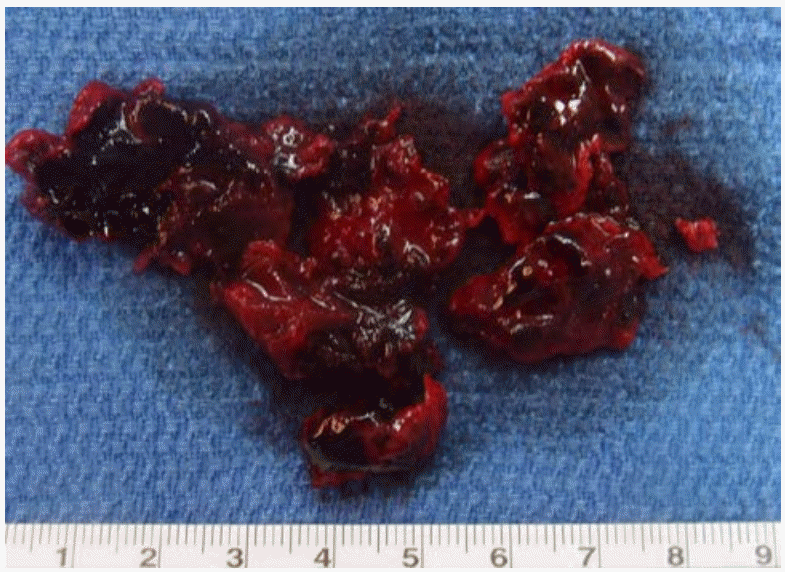
The LA was approached by extending an incision from the RAtomy for Maze to the posterior RA appendage and opening the LA roof. The posterior wall of the LA roof was swollen, and it almost filled the entire left atrium. However, no tear or puncture was found on the internal wall of the left atrium, and the wall color was normal. Needle aspiration of the posterior wall of the swollen LA was performed with no initial result. However, when the opening was enlarged, an unorganized hematoma was suctioned out. A 4-cm incision was made at the opening, and a hematoma was evacuated using a suction tip and forceps until the outer wall of the LA could be visualized (Figure 3). The patient recovered without any immediate postoperative complications.
Over a 1-year follow-up, no recurrences of AT or AF were noted, and the patient maintained sinus rhythm.
Discussion
RFCA is typically performed through a percutaneous approach under fluoroscopic guidance. The incidence of systemic thromboembolic complications associated with RFCA in the left heart has been reported to be as high as 1-5%, but bleeding complications, such as intramural hematoma, are rare [5-7]. An intramural hematoma can be diagnosed using various imaging modalities, including echocardiography, cardiac computed tomography, and cardiac magnetic resonance imaging, which show a non-enhancing mass within the wall of the atrium.
The findings in the present case suggest that optimal periprocedural anticoagulation with heparin might reduce the risk of procedure related thromboembolic complications, but physicians should always pay attention to the risk of bleeding. Post-procedural imaging studies and short-term follow-up could help in the early detection and prevention of fatal bleeding complications.


