|
|
International Journal of Arrhythmia 2014;15(3): 62-68.
|
 |
| ECG & EP CASES |
Anatomical Obstacles to
Catheter Ablation for
Atrioventricular Nodal
Reentrant Tachycardia |
 
|
|
|
|
Introduction
Radiofrequency catheter ablation (RFCA) is
the first choice of treatment for symptomatic
AVNRT.1 However, its use in patients with anatomic
variations can be complicated. Here, we
present two cases of catheter ablation for AVNRT
in patients with anatomic variations: an RA
septal diverticulum, and lung-disease-induced
heart distortion, respectively.
Case 1
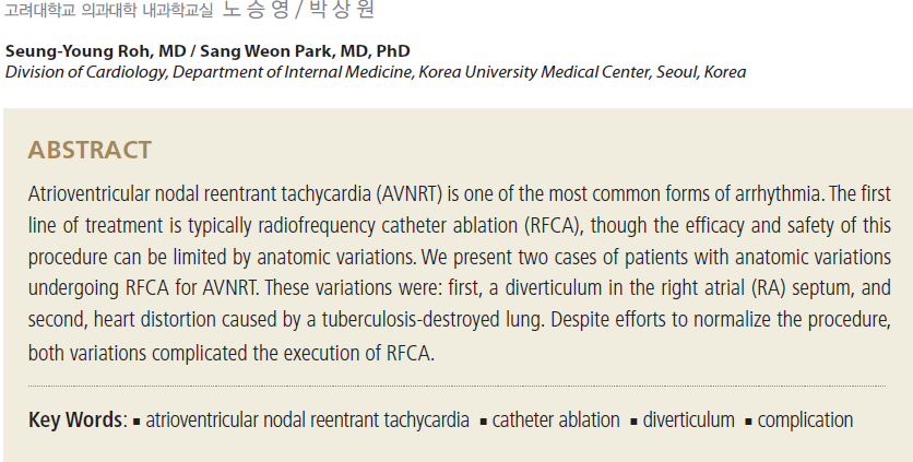
A 22-year-old woman presented with paroxysmal
palpitation. Electrocardiography (ECG)
revealed narrow QRS tachycardia with a pulse
rate of 160 beats/min during palpitation (Figure
1). The patient’s blood pressure was 110/80 mmHg
during tachycardia. QRS rhythm was regular and
pseudo R’ wave was observed in the precordial
lead from V1 to V3. Sinus rhythm was restored
following rapid administration of intravenous
adenosine (6 mg). The patient had no history of
disease or operations. A transthoracic echocardiogram
(TTE) showed normal left ventricular
ejection fraction (60%) and no structural abnormalities.
For electrophysiological (EP) investigation,
a 2-mm and a 4-mm quadripolar catheter
were used to record His and right ventricular (RV)
activity, respectively. Unfortunately, placement of a duodecapolar catheter into the coronary sinus
(CS) failed as it could not be advanced into
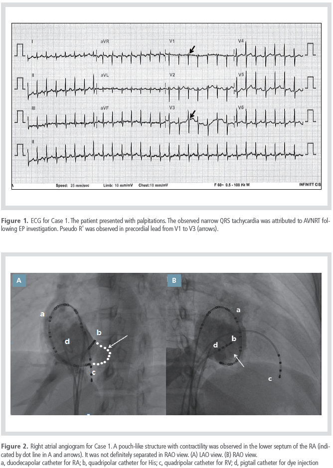
the CS ostium. A right atrial (RA) angiogram was
performed for structural analysis (Figure 2). A
pouch-like structure was observed in the lower
septum of the RA, near the CS ostium. As this
structure exhibited contractility, it was diagnosed
as a diverticulum, rather than a septal aneurysm.
A CS angiogram revealed no association between
the diverticulum and the CS (Figure 3). Attempts
to place the duodecapolar catheter in the CS were
impeded by the diverticulum. An EP study was
subsequently performed using a duodecapolar
catheter positioned at the RA. Tachycardia
was induced after an atrio-His (AH) jump, and
atrioventricular and ventriculoatrial conduction
exhibited decremental properties. Clinical tachycardia
was attributed to slow-fast AVNRT after
differential diagnostic maneuvers. A deflectable
ablation catheter with a 4-mm tip was positioned
at the anterior margin of the CS to ablate the
slow pathway. The ablation catheter was found
to be unstable yet it was easily moved up and
down at the margin of the septal diverticulum. As
a result, successful RFCA was only achieved after
a considerable time interval.

Case 2
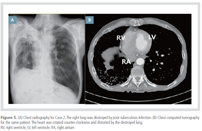
A 71-year-old man with a tuberculosis-destroyed
lung presented with palpitation and dyspnea.
Electrocardiography (ECG) revealed narrow-
QRS tachycardia with a short RP interval
and a pulse rate of 170 beats/min during palpitation
(Figure 4). The patient’s blood pressure was
100/70 mmHg at the time of recording, and QRS
rhythm was regular. Sinus rhythm was restored
following rapid administration of intravenous adenosine
(6 mg). The patient had diabetes mellitus,
hypertension, and a history of pulmonary tuberculosis. A TTE showed preserved left ventricular
ejection fraction (55%) and no structural
abnormality. Chest radiography and chest computed
tomography showed a severely distorted
lung (Figure 5), and counter-clockwise rotation
of the heart. In RA angiography, the RA exhibited
erect morphology. An EP investigation was subsequently
performed using a 2-mm and a 4-mm
quadripolar catheter to record His and RV activity,
respectively. A duodecapolar catheter was
positioned at the CS and the RA. Clinical tachycardia
was attributed to slow-fast AVNRT on the
basis of EP investigation. Due to the high risk of
atrioventricular (AV) block, owing to the patient’s
advanced age and distorted heart structure, the
ablation focus was carefully considered. First,
the lowest level for detection of His potential was
identified (Figure 6A, B). Next, a posterior approach
was taken, via the middle or posterior
septal region near the CS ostium (Figure 6D). His
potential was not observed on the electrogram of
the ablation catheter (Figure 6C). Energy delivery
resulted in successful induction of junctional
rhythm, though ablation was immediately aborted
on observing ventriculoatrial (VA) conduction
block some seconds later. A high degree of AV
block with concurrent hypotension occurred. The
AV block was initially sustained but eventually
recovered after eight hours; the PR interval normalized
after two weeks.

Discussion
AVNRT is one of the most common tachyarrhythmias,
and can be treated by catheter ablation.
This can be hazardous when the slow pathway
is in close proximity to the normal conduction
system. Thus, a clear understanding of cardiac
anatomy is essential before AVNRT ablation.
We have reported two complicated AVNRT
cases related to right heart anatomic abnormalities.
In the first case, an RA septal diverticulum
compromised the positioning and stability of the
catheter. Binder et al. analyzed 103 cases of congenital
malformations of the RA and the CS.2 Of
the 103 cases studied, 13 were associated with an
RA single diverticulum and these were predominantly
asymptomatic. The presentation of symptoms
such as supraventricular tachycardia was
frequently induced by arrhythmia.
We present the first reported case of a single
diverticulum in the RA septum. Previous studies
have reported cases of RA diverticula predominantly
localized to the RA free wall or the CS.2-7
The RA septal diverticulum described in this case
was separated from the CS, as demonstrated by
the angiogram. Because the diverticulum exhibited
contractility consistent with the heartbeat,
we ruled out the alternative diagnosis of septal
aneurysm, in which contractility would not be
observed.8
Acquired anatomic distortions can also interfere
with RFCA for AVNRT. In the second case,
safety was ensured by using numerous methods:
(1) RA angiogram, (2) confirmation of the lowest
point for detection of His potential, (3) a posterior
approach near the CS ostium, and (4) vigilant
observation of VA conduction. A contemporary
transient high degree AV block was nevertheless
seen to occur.
For effective and safe catheter ablation in patients
with anatomic obstacles, an overview of
the precise anatomy is critical. Angiograms and
careful mapping can facilitate the identification of
anatomic variants, and can confirm precise catheter
positioning.
Conclusion
We have reported two difficult AVNRT cases related
to right heart anatomic variation: the first,
an RA septal aneurysm, and the second, heart
distortion due to tuberculosis-destroyed lung.
Anatomic obstacles can compromise successful
catheter ablation for AVNRT.
References
- Katritsis DG, Camm AJ. Atrioventricular nodal reentrant tachycardia.
Circulation. 2010;122:831-840.
- Binder TM, Rosenhek R, Frank H, Gwechenberger M, Maurer G, Baumgartner H. Congenital malformations of the right atrium and the coronary sinus: an analysis based on 103 cases reported in the literature and two additional cases.
Chest. 2000;117:1740-1748.
- Morrow AG, Behrendt DM. Congenital aneurysm (diverticulum) of the right atrium. Clinical manifestations and results of operative treatment.
Circulation. 1968;38:124-128.
- Di Segni E, Siegal A, Katzenstein M. Congenital diverticulum of the heart arising from the coronary sinus.
Br Heart J. 1986;56:380-384.
- Pastor BH, Forte AL. Idiopathic enlargement of the right atrium.
Am J Cardiol. 1961;8:513-518.
- Morishita Y, Kawashima S, Shimokawa S, Taira A, Kawagoe H, Nakamura K. Multiple diverticula of the right atrium.
Am Heart J. 1990;120:1225-1227.
- Sheldon WC, Johnson CD, Favaloro RG. Idiopathic enlargement of the right atrium. Report of four cases.
Am J Cardiol. 1969;23:278-284.
- Mugge A, Daniel WG, Angermann C, Spes C, Khandheria BK, Kronzon I, Freedberg RS, Keren A, Denning K, Engberding R, Sutherland GR, Vered Z, Erbel R, Visser CA, Lindert O, Hausmann D, Wenzlaff P. Atrial septal aneurysm in adult patients. A multicenter study using transthoracic and transesophageal echocardiography.
Circulation. 1995;91:2785-2792.
|
|
|
|


