International Journal of Arrhythmia 2012;13(4): 37-41.
Untitled Document
ECG & EP CASES
Spontaneous Intramural Hematoma of the Small Bowel after Oral Anticoagulation Therapy
대구가톨릭대학교 의과대학 내과학교실 이 영 수
Young-Soo Lee, MD, PhD, Cardiology Division, Department of Internal Medicine, Daegu Catholic University College of Medicine, Daegu, Korea
Introduction
Long-term oral anticoagulation (OAC) therapy
has been recommended for stroke prevention in
patients with atrial fibrillation (AF) and is
prescribed according to scoring systems such as
CHADS2 or CHA2DS2-VASc. On the other hand,
OAC increases bleeding risk on excessive
accumulation of the oral anticoagulant, which is
associated with various hemorrhagic complications such as hematuria, gastrointestinal bleeding,
intracerebral hemorrhage, soft tissue hematomas,
epistaxis, and retroperitoneal hematomas.
Intramural hematoma of the small bowel is a rare
complication of the use of OAC therapy. The
condition usually presents as abdominal pain,
which is frequently accompanied by nausea and
vomiting. A history of OAC therapy use with
prolonged international normalized ratios (INRs)
should be considered in the diagnosis of patients
presenting with abdominal pain.
We present a case wherein an AF patient
developed a spontaneous intramural hematoma of
the small bowel after OAC therapy.
Case
A 67-year-old woman presented to our
institution with abrupt-onset abdominal pain. She
was taking aspirin and clopidogrel for 8 years
because of cerebellar infarction and therapy
involving an oral anticoagulant (warfarin 2 mg/day)
and amiodarone (200 mg/day) for 1 year because of
atrial fibrillation. She did not have a history of
trauma. Her blood pressure was 135/85 mmHg.
Physical examination showed abdominal distension
with tenderness and that bowel sounds had
decreased. The following laboratory test results
were obtained: hemoglobin level 11.8 g/dL, white
blood cell count 11,020/mm3, and platelet count
319,000/mm3. The coagulation test showed a
prothrombin time of 60.4 s and an INR of 5.26. The
creatinine level was slightly elevated (1.6 g/dL). An
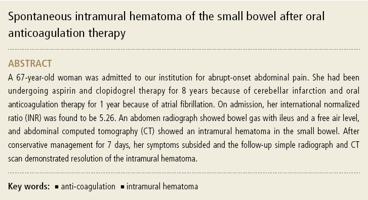
abdominal radiograph showed ileus and air-fluid
levels indicating intestinal obstruction (Figure 1A
and 1B). Computed tomography (CT) demonstrated mural thickening and an intramural hematoma in
the small bowel (Figure 2A and 2B). OAC was
therapy stopped immediately and vitamin K was
given intravenously. Furthermore, parenteral
nutrition was initiated for bowel rest. Consequently,
the INR value returned within normal range, and
the bowel sounds increased. After 7 days, the
follow-up radiograph and CT scan demonstrated
resolution of the previous ileus and mural
thickening, respectively (Figures 3 and 4). The
patient then began oral nutrition and was
discharged when she passed yellowish stools. She
has been free of symptoms and is only undergoing
aspirin treatment as an outpatient.
Discussion
Long-term OAC therapy has been recommended
for preventing stroke in patients with AF,
according to scoring systems. However, OAC
increases bleeding risk on excessive accumulation of the anticoagulant. Because of the narrow
therapeutic range of OAC therapy, patients
undergo capillary blood sampling for measuring
prothrombin time (PT). The PT is standardized as
the INR with a target range of 2.0~3.0. With OAC
therapy, the annual risk of major bleeding
increases significantly to 0.3%.1 The most severe
bleeding complication is intracranial hemorrhage.
INR values of >4.0 have been known to increase
the risk of major hemorrhage.2 Furthermore, some
schemes have been reported for predicting bleeding
risk. Gage, et al.3 presented the HEMORRHAGES
score, which considers the following factors:
liver/renal disease, alcohol abuse, malignancy, age
>75 years, low platelet count or function,
rebleeding risk, uncontrolled hypertension, anemia,
genetic factors (CYP2C9), and risk of fall or stroke,
with 1 point for each risk factor present or 2 points
for a previous bleed. The HAS-BLED score has been
recently reported to allow assessment of bleeding
risk for patients with AF in the SPORTIF cohort.4,5
The HAS-BLED score includes hypertension,
abnormal renal/liver function, stroke, bleeding history or predisposition, labile INR (<60% of the
time in the therapeutic range), elderly age (age >75
years), and concomitant drugs and alcohol. A score
of more than 2 for the HAS-BLED scoring system is
considered to indicate a high risk of major bleeding.
Spontaneous intestinal intramural hematoma is
an uncommon complication of anticoagulation. The
incidence of spontaneous intramural hematoma
is reported to be 1 in 2,500 patients using
anticoagulation therapy.6 The jejunum is commonly
involved, followed by the ileum and the duodenum.6
The clinical manifestations vary from vague
abdominal pain, nausea, vomiting, acute abdomen
or intestinal obstruction, and gastrointestinal
bleeding.7 The management approach involves
medical treatment, discontinuation of anticoagulant
drugs, bowel rest, correction of PT with intravenous
vitamin K, and fresh frozen plasma.7 If correctly
diagnosed pre-operatively, conservative management
with restoration of coagulation parameters leads to
a satisfactory recovery in most cases. Surgical
intervention is indicated only if there is significant
intramural hemorrhage, bowel perforation, ischemia, or peritonitis.8
Taken together, intramural hematoma is an
uncommon hemorrhagic complication of long-term
anticoagulation therapy and should be considered
in patients presenting with acute abdomen pain.
Early diagnosis enables treatment of most patients
without an invasive operation.