이식형 심장 사건 기록기의 임상적 역할
Abstract
Initially, an implantable loop recorder (ILR) was introduced to find the cause of unexplained syncope, palpitation, and dizziness. It is now considered as a test of choice when repeated Holter recordings, more-prolonged external event loop recordings, head-up tilt test, or electrophysiological study fails to reveal the cause of recurrent syncope. However, with the help of technical advancement such as automated QRS morphology analysis, auto-adjusting sensing threshold, improved noise rejection, increased battery longevity, and remote monitoring function, the ILR has begun to play an important role in the diagnosis of life-threatening ventricular tachyarrhythmias as well. In addition, ILR monitoring shows clear usefulness in detecting atrial fibrillations after cryptogenic stroke, in differentiating true syncope from epilepsy, and in assessing the recurrence rate of atrial fibrillation after radiofrequency ablation. Overall, a smarter ILR will make possible earlier diagnosis, enhance diagnostic accuracy, and improve quality of life of patients with unexplained syncope, palpitation, non-syncopal transient loss of consciousness, and stroke.
Key words: Syncope; Atrial Fibrillation; Ventricular Tachycardia; Stroke
서론
실신은 매우 흔한 질환임에도 불구하고 정확한 원인을 진단하는 것이 쉽지 않다. 기립경사 테이블 검사, 심장 초음파 및 관상동맥 조영술, 심장 전기생리학 검사, 뇌영상 및 뇌파검사 등의 신경과적인 검사를 포함한 종합적인 평가를 하더라도 실신의 원인이 밝혀지지 않는 경우가 약 1/3 정도나 되는 것으로 알려져 있다[ 1, 2]. 그런데 원인불명의 실신을 경험한 환자는 실신을 경험하지 않은 환자에 비해 예후가 나쁜 것으로 알려져 있어, 원인을 정확하게 진단하여 바른 치료를 제공하는 것이 매우 중요한 과제이다[ 3]. 이러한 원인불명의 실신이나 일시적 의식소실 환자에서 부정맥으로 인한 원인을 밝히는 데에 매우 유용한 진단 방법이 바로 이식형 심장 사건 기록기(implantable loop recorder, ILR)이다[ 4]. 최근에는 기술적인 발전에 힘입어 ILR의 수명이 길어지고, 진단의 정확도가 높아지고 있다. 따라서 기존에 주로 사용되던 원인불명의 실신 외에, 심방세동, 원인불명의 뇌졸중(cryptogenic stroke), 간질(epilepsy)과 같은 질환에서도 응용 범위가 점차 확대되고 있다.
ILR의 종류, 기능, 이식
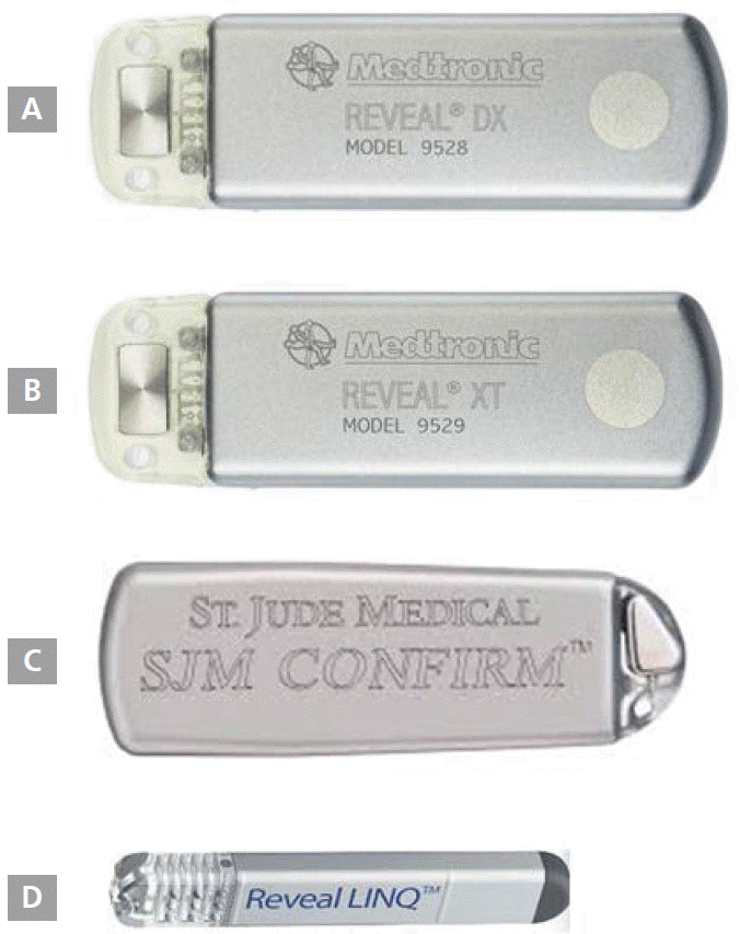
ILR은 USB 메모리 stick과 유사하게 납작한 직사각형의 작은 상자 모양을 하고 있으며, single-lead 심전도를 기록하고 저장할 수 있는 기능을 갖고 있다( Figure 1). 일정한 범위의 서맥이나 빈맥이 발생하면 자동으로 심전도를 저장하도록 프로그래밍할 수 있고, 환자가 증상을 느낄 때 추가적으로 심전도를 기록하도록 ILR을 활성화시킬 수도 있다. 현재 일반적으로 사용되고 있는 ILR의 제품별 사양은 Table 1에 정리하였다. ILR은 일반적으로 좌흉골연 부위(left parasternal region)에 세로로 이식한다. 최근에는 외래에서 피하주사하듯이 간단하게 이식이 가능할 정도로 매우 작아진 injectable ILR(Medtronic Reveal LINQ™)이 개발되었으며, 조만간 국내에도 도입되어 환자에게 보다 많은 편의를 제공할 것으로 기대되고 있다( Figure 1).
원인불명의 실신에서 ILR의 유용성
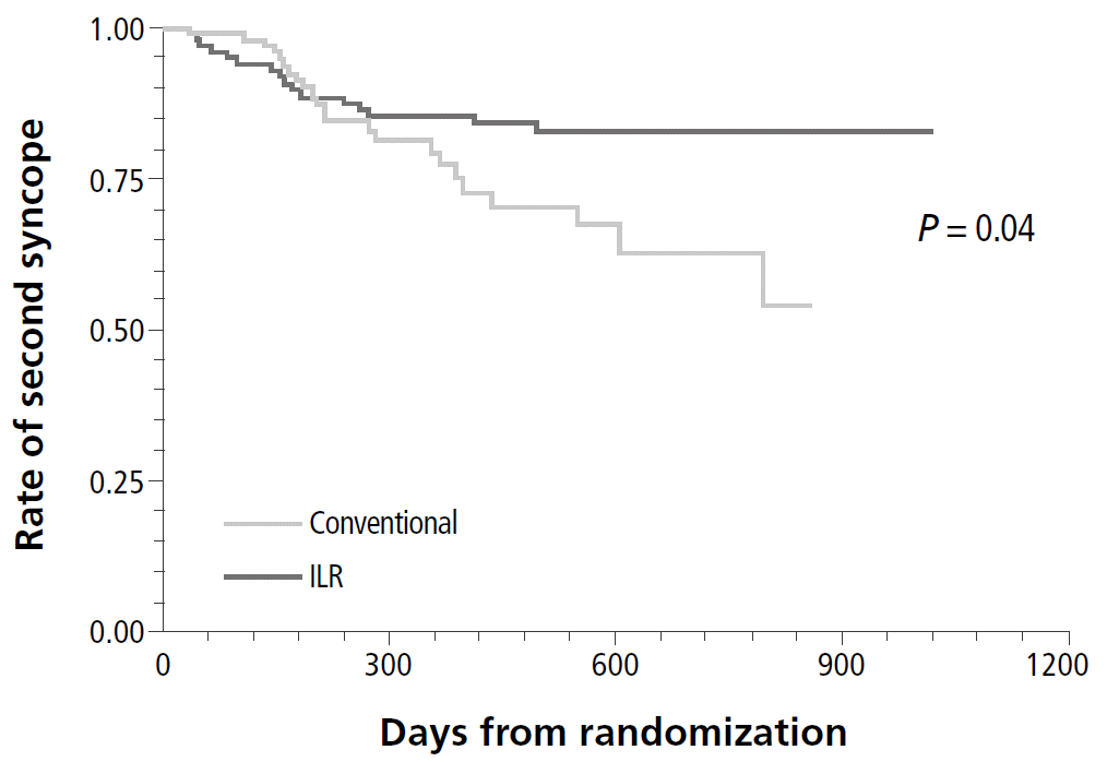
ILR은 기존의 진단법에 비해 원인불명의 실신이나 의식소실의 원인에 대한 진단율이 높다. 홀터모니터링 검사, 비이식형 사건 기록기(external loop recorder), 전기생리학 검사 및 신경과적 영상기법 등은 대부분 20% 미만의 낮은 진단율을 보이고 있고, 기립경사 테이블 검사의 진단율이 상대적으로 높게 보고되기도 하나 일관되지 못한 성적을 보이고 있다(19-61%) [ 1, 2]. 이에 반해 ILR의 진단율은 50-80% 정도로 높게 보고되는 경우가 많다[ 5, 6]. 따라서 2009년에 발표된 European Society of Cardiology (ESC) 임상지침에서는 원인불명의 실신이 반복되는 환자에게 ILR을 조기에 사용할 것을 class I으로 권고하고 있다[ 7]. ILR로 증명된 실신의 부정맥 원인으로는 동기능 부전이나 고도 방실 차단과 같은 서맥성 부정맥이 가장 흔하다[ 5, 8]. 우리나라에서는 2006년부터 ILR이 이식되기 시작하여 2013년까지 75명의 환자가 ILR 이식술을 받았으며, ILR을 통해 진단된 원인 중 서맥성 부정맥이 약 2/3를 차지하여 외국과 유사한 경향을 보여주었다. 또한 심장 돌연사를 일으킬 수 있는 심실빈맥이나 심실세동으로 진단된 경우도 30% 정도로 높게 나타나 심실빈맥의 진단에 있어서도 ILR이 유용할 수 있음을 시사하였다(2014년 대한순환기관련학회 춘계통합학술대회). 또한 영국에서 원인불명의 환자를 대상으로 진행된 EaSyAS (Eastbourne Syncope Assessment Study) 연구는 기존 진단법에 근거한 치료에 비하여 ILR 진단에 근거한 치료 결과 실신의 재발이 현저하게 감소함을 보고하여 ILR이 보다 정확한 치료 방법을 결정하는 데에 매우 중요한 역할을 하였음을 보여주었다( Figure 2) [ 9].
심방세동 진단을 위한 ILR
ILR은 심방세동이 의심되는 경우에 오랜 기간 동안 심전도 모니터링을 가능하게 해주어 심방세동 진단에 도움을 줄 수 있다. 좌심실 기능이 저하된 초기 급성 심근경색 환자 300여 명을 대상으로 ILR을 이식하고 진행된 CARISMA (Cardiac Arrhythmias and Risk Stratification After Myocardial Infarction) 임상 연구 결과 28%의 환자에서 심방세동이 새롭게 발견되었다. 또한 ILR은 심방세동 전극도자 절제술 후에 재발을 평가하는 방법으로 사용되기도 한다[ 10]. ILR은 재발 유무 외에 심방세동의 burden도 측정할 수 있다는 장점이 있다. 최근에 Mittal 등이 발표한 흥미로운 연구 결과에 따르면 심방세동이 증명되지 않고 심방조동만 있었던 20명의 환자를 cavotricuspid isthmus (CTI) 절제술 후 평균 1년 정도 경과를 관찰한 결과 11명(55%)의 환자에서 심방세동이 관찰되었다[ 11]. 따라서 심방조동만이 관찰되어 CTI 절제술로 정상 동율동이 잘 유지되는 것으로 보이는 환자들도 CHA 2DS 2-VASc 점수가 높다면 항응고 치료를 지속해야 할지에 대한 추가적인 연구가 필요할 것으로 생각된다.
원인불명의 뇌졸중 환자와 ILR
여러 가지 자세한 검사를 해도 원인이 밝혀지지 않은 원인불명의 뇌졸중은 전체 뇌경색 환자의 약 1/3을 차지하는 것으로 알려져 있다. 이러한 경우에 심전도 모니터링을 한 달 가까이 연장해 보면 약 20-23%의 환자에서 발작성 심방세동이 관찰되기 때문에, 발작성 심방세동은 원인불명 뇌경색의 중요한 원인으로 주목받고 있다[ 12, 13]. 따라서 ILR은 원인불명 뇌경색 환자들에서 원인규명, 즉 심방세동 발견에 중요한 역할을 할 것으로 기대되고 있다. 2013년 Etgen 등이 발표한 결과에 따르면 393명의 뇌경색 환자 중 16.5%인 65명이 원인불명의 뇌경색이었고, 이들 중 22명에서 ILR 삽입술을 시행할 수 있었으며, 이들을 대상으로 1년간 관찰한 결과 27.3%인 6명의 환자에서 심방세동을 발견할 수 있었다[ 14].
ILR의 기술적 발전과 심실빈맥의 발견
ILR 사용 초기에는 주로 원인불명의 실신 환자에서 서맥성 부정맥을 발견하기 위해 사용되었다. 앞에서도 이미 설명했듯이 서맥성 부정맥이 원인불명의 실신 환자에서 가장 흔한 심장 맥박의 이상소견이지만, 약 10%에서는 심실 부정맥이 발견된다는 보고도 있다[ 15]. ILR을 이용한 심전도 분석은 single channel만 이용 가능하다는 점, signal undersensing (특히 P-wave), T-wave oversensing, noise oversensing 등의 여러 가지 단점이 있다. 따라서 심실빈맥을 진단하는 면에서 ILR의 유용성이 다소 떨어질 수 있다는 우려가 있다. 하지만 최근 automated QRS wave morphology analysis, auto-adjusting sensing threshold, RR interval regularity analysis, pattern of heart rate increase analysis 등의 알고리즘이 도입되어 상심실성 빈맥과 심실성 빈맥의 감별 정확도가 높아지게 되었다. ICD 삽입(implantation) 당시 함께 ILR을 삽입하고, 심실빈맥이나 심실세동을 유발한 한 연구 결과 ILR은 심실 부정맥을 95-99% 정도로 정확하게 진단하였다[ 16]. 또한 Ergul Y 등은 12명의 원인불명 실신의 소아 환자들에게 ILR을 이식하고, 평균 20개월을 추적 검사한 결과 5명(42%)의 환자에서 Torsade de pointes, 심실세동, 심실빈맥이 관찰되어[ 17] ILR이 치명적인 심실 부정맥을 조기에 발견하는 데 도움이 될 수 있음을 시사하였다.
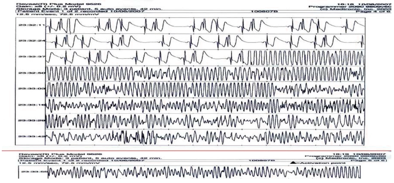
Figure 3는 원인불명 실신의 원인이 심실세동이었음이 ILR로 잘 증명된 국내 증례이다[ 18]. 반복되는 실신과 급성 심장사의 가족력(부친)을 주소로 입원한 27세 남성 환자로 심장 초음파, 기립경사 테이블 검사, 심장 전기 생리학 검사 및 뇌파 검사에서는 특이 소견이 없었다. ILR 시술 후 5개월째 밤에 자다가 갑자기 경련성 실신이 발생하였고, 당시 ILR 기록 상 심실빈맥 및 심실세동이 90초간 기록되어 ILR을 제거하고 삽입형 심실 제세동기를 시술하였다.
간질과 ILR
실신이 사지의 이상한 움직임을 동반할 때(convulsive reflex syncope) 간질과의 감별이 어려울 수 있다. 기존의 연구에 따르면 간질 환자의 13-42%가 오진인 것으로 밝혀졌고, 이 중 대부분이 실신이 원인이었다[ 19, 20]. 이러한 경우 ILR은 실신 당시 심장 박동의 변화뿐 아니라 tonic-seizure에 동반되는 근전위(myopotential [muscle artifacts])를 보여줄 수 있기 때문에 이 두 가지를 보다 정확히 감별하는 데에 매우 유용하게 쓰일 수 있다( Figure 4) [ 21]. 2012년 Europace에 발표된 전향적 연구인 REVISE(Reveal in the Investigation of Syncope and Epilepsy) 연구 결과에 따르면 기존에 간질의 진단이 다소 불분명했던 103명의 환자에게 ILR을 이식하고, 평균 2.4년을 경과 관찰하였을 때 21%인 22명의 환자에서 경련 움직임(convulsive movement)을 동반한 심한 서맥(bradyarrhythmia or asystole)이 관찰되어 진단명이 바뀌게 되었다. 이들은 간질약을 끊고 인공심장 박동기 이식을 한 후에 증상이 더 이상 발생하지 않았다[ 21].
결론
ILR은 원인불명의 실신 환자에서 서맥성 부정맥을 조기에 발견하는 데에 최선의 검사일 뿐만 아니라 기술적인 발전에 힘입어 치명적인 심실성 부정맥의 발견에도 도움이 되고 있다. 최근에는 점차 그 사용 영역이 확대되고 있어 원인불명의 심계항진, 심방세동 및 심방조동 치료 후에 심방세동의 발생의 추적관찰, 원인불명의 뇌경색 환자에서 원인 질환 규명, 간질과 실신의 감별 등에도 유용하게 사용되고 있다. 특히 무선 통신 기술 도입으로 인한 remote 모니터링 기능이 추가되어 향후 조기 진단, 조기 예방, 조기 치료가 더욱 활발해지고, 따라서 환자의 삶의 질을 크게 향상시킬 것으로 기대되고 있다.
References
1. Kang GH, Oh JH, Kim JS, On YK, Song HG, Jo IJ, Kim SJ, Bae SJ, Shin TG. Diagnostic patterns in the evaluation of patients presenting with syncope at the emergency or outpatient department. Yonsei Med J 2012; 53: 517-523.    2. Pires LA, Ganji JR, Jarandila R, Steele R. Diagnostic patterns and temporal trends in the evaluation of adult patients hospitalized with syncope. Arch Intern Med 2001; 161: 1889-1895.   3. Soteriades ES, Evans JC, Larson MG, Chen MH, Chen L, Benjamin EJ, Levy D. Incidence and prognosis of syncope. N Engl J Med 2002; 347: 878-885.  4. Edvardsson N, Frykman V, van Mechelen R, Mitro P, Mohii-Oskarsson A, Pasquie JL, Ramanna H, Schwertfeger F, Ventura R, Voulgaraki D, Garutti C, Stolt P, Linker NJ; PICTURE Study Investigators. Use of an implantable loop recorder to increase the diagnostic yield in unexplained syncope: results from the PICTURE registry. Europace 2011; 13: 262-269.    5. Kang GH, Oh JH, Chun WJ, Park YH, Song BG, Kim JS, On YK, Park SJ, Huh J. Usefulness of an implantable loop recorder in patients with syncope of an unknown cause. Yonsei Med J 2013; 54: 590-595.    6. Furukawa T, Maggi R, Bertolone C, Fontana D, Brignole M. Additional diagnostic value of very prolonged observation by implantable loop recorder in patients with unexplained syncope. J Cardiovasc Electrophysiol 2012; 23: 67-71.   7. Task Force for the Diagnosis and Management of Syncope; European Society of Cardiology (ESC); European Heart Rhythm Association (EHRA); Heart Failure Association (HFA); Heart Rhythm Society (HRS), Moya A, Sutton R, Ammirati F, Blanc JJ, Brignole M, Dahm JB, Deharo JC, Gajek J, Gjesdal K, Krahn A, Massin M, Pepi M, Pezawas T, Ruiz Granell R, Sarasin F, Ungar A, van Dijk JG, Walma EP, Wieling W. Guidelines for the diagnosis and management of syncope (version 2009). Eur Heart J 2009; 30: 2631-2671.    8. Boersma L, Mont L, Sionis A, Garcia E, Brugada J. Value of the implantable loop recorder for the management of patients with unexplained syncope. Europace 2004; 6: 70-76.   9. Farwell DJ, Freemantle N, Sulke N. The clinical impact of implantable loop recorders in patients with syncope. Eur Heart J 2006; 27: 351-356.   10. Verma A, Champagne J, Sapp J, Essebag V, Novak P, Skanes A, Morillo CA, Khaykin Y, Birnie D. Discerning the incidence of symptomatic and asymptomatic episodes of atrial fibrillation before and after catheter ablation (DISCERN AF): a prospective, multicenter study. JAMA Intern Med 2013; 173: 149-156.   11. Mittal S, Pokushalov E, Romanov A, Ferrara M, Arshad A, Musat D, Preminger M, Sichrovsky T, Steinberg JS. Long-term ECG monitoring using an implantable loop recorder for the detection of atrial fibrillation after cavotricuspid isthmus ablation in patients with atrial flutter. Heart Rhythm 2013; 10: 1598-1604.   12. Elijovich L, Josephson SA, Fung GL, Smith WS. Intermittent atrial fibrillation may account for a large proportion of otherwise cryptogenic stroke: a study of 30-day cardiac event monitors. J Stroke Cerebrovasc Dis 2009; 18: 185-189.   13. Tayal AH, Tian M, Kelly KM, Jones SC, Wright DG, Singh D, Jarouse J, Brillman J, Murali S, Gupta R. Atrial fibrillation detected by mobile cardiac outpatient telemetry in cryptogenic TIA or stroke. Neurology 2008; 71: 1696-1701.   14. Etgen T, Hochreiter M, Mundel M, Freudenberger T. Insertable cardiac event recorder in detection of atrial fibrillation after cryptogenic stroke: an audit report. Stroke 2013; 44: 2007-2009.   15. Nierop PR, van Mechelen R, van Elsacker A, Luijten RH, Elhendy A. Heart rhythm during syncope and presyncope: results of implantable loop recorders. Pacing Clin Electrophysiol 2000; 23: 1532-1538.   16. Volosin K, Stadler RW, Wyszynski R, Kirchhof P. Tachycardia detection performance of implantable loop recorders: results from a large 'real-life' patient cohort and patients with induced ventricular arrhythmias. Europace 2013; 15: 1215-1222.   17. Ergul Y, Tanidir IC, Ozyilmaz I, Akdeniz C, Tuzcu V. Evaluation Rhythm Problems in Unexplained Syncope Etiology with Implantable Loop Recorder. Pediatr Int 2015; 57: 359-366.   18. Shin DH, Kim JS, Park JW, Yim HR, Kim JH, Lee SM, Kim KS, Lee CH, Jung DC, On YK, Kim DK, Lee SH, Hong KP, Park JE. The Use of an Implantable Loop Recorder in Patients with Syncope of Unknown Origin. Korean Circ J 2008; 38: 205-211.  19. Zaidi A, Clough P, Cooper P, Scheepers B, Fitzpatrick A. Misdiagnosis of epilepsy: many seizure like attacks have a cardiovascular cause. J Am Coll Cardiol 2000; 36: 181-184.   20. Josephson CB, Rahey S, Sadler RM. Neurocardiogenic syncope: frequency and consequences of its misdiagnosis as epilepsy. Can J Neurol Sci 2007; 34: 221-224.   21. Petkar S, Hamid T, Iddon P, Clifford A, Rice N, Claire R, McKee D, Curtis N, Cooper PN, Fitzpatrick AP. Prolonged implantable electrocardiographic monitoring indicates a high rate of misdiagnosis of epilepsy-REVISE study. Europace 2012; 14: 1653-1660.  
Figure 1.
Several types of implantable loop recorder; Medtronic Reveal® DX (A), Medtronic Reveal®XT (B), St. Jude Confirm™ Implantable Cardiac Monitor (ICM, C), and Medtronic Reveal LINQ™ (D). 
Figure 2.
Time to second syncope recurrence. Implantable loop recorder patients achieved a longer time to second syncope, suggesting a more appropriate treatment. ILR, implantable loop recorder. 
Figure 3.
Ventricular fibrillation recorded by using implantable loop recorder during sleep in a 27 year-old male patient with unexplained recurrent syncopal episodes. 
Figure 4.
Electrocardiograms recorded in implantable loop recorder during tonic-clonic seizure. (A) Continuous muscle artifacts can be seen, indicating tonus. (B) Discrete muscle artifacts of the clonus. (C) The clonic artifacts attenuated and the electrocardiographic complexes becoming visible again as the seizure began to fade. 
Table 1.
Several types of implantable loop recorder
|
Medtronic Reveal® DX |
Medtronic Reveal® XT |
St. Jude Confirm |
|
Size |
62 x 19 x 8 mm |
62 x 19 x 8 mm |
56.3 x 18.5 x 8 mm |
|
9 mL |
9 mL |
6.5 mL |
|
Weight |
15 g |
15 g |
12 g |
|
Asystole, |
Asystole, |
Asystole, |
|
Arrhythmia |
bradyarrhythmia VT/fast |
bradyarrhythmia VT/fast |
bradyarrhythmia VT/fast |
|
Detection |
VT |
VT, AT/AF, |
VT |
|
|
AT/AF burden, HRV |
|
|
Longevity |
3 years |
3 years |
3 years |
|
Event storage |
49.5 minutes |
49.5 minutes |
48 minutes |
|
MRI labeling |
MR conditional |
MR conditional |
MR conditional |
|
Availability (Korea) |
Yes |
No |
Yes |
|
|


