Introduction
Premature ventricular contractions (PVCs) or ventricular tachycardia (VT), with QRS morphologies of left bundle branch block (LBBB) and inferior axis can be ablated successfully from the right ventricular outflow tract (RVOT). However, it is not uncommon that PVCs or VT assumed to originate from the RVOT actually arise from the aortic root because of the proximity of the RVOT to the aortic root. Spatially, the aortic valve lies to the right and posterior to the RVOT [1].
We present a patient with right coronary cusp origin PVCs, exhibiting two distinct QRS morphologies with LBBB pattern.
Case
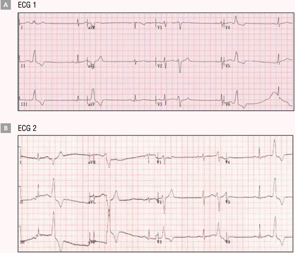
A 52-year-old woman presented at the emergency center in our hospital for syncope. She had been diagnosed with RVOT VT at a private clinic 15 years previously, and amiodarone was prescribed for VT. However, amiodarone induced thyrotoxicosis, and as a result, sotalol was prescribed as an alternative. Several years later, sotalol became unavailable in Korea, requiring the subsequent prescription of metoprolol. In our hospital, verapamil or flecainide was prescribed for PVCs and VT but was ineffective in alleviating her symptoms. In visits to our clinic, she complained of palpitations and fainting, while her electrocardiogram (ECG) revealed frequent PVCs characterized by LBBB pattern and inferior axis. The majority of PVCs presented with positive QRS complex morphology in lead I, but a small number showed a negative QRS complex in lead I (Figure 1). Ultimately, radiofrequency catheter ablation was required to eliminate PVCs and VT.
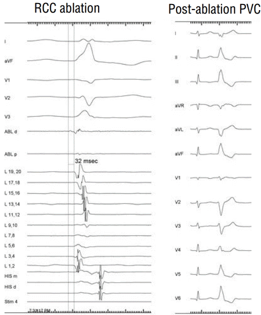
At baseline ECG, QRS morphology of monomorphic PVCs (PVCs 1) exhibited a LBBB pattern and inferior axis. It was suspected that observed PVCs were originating from the RVOT, and consequently activation and pace mapping was performed at the RVOT. During mapping, other PVCs (PVCs 2 and PVCs 3) were observed. PVCs 3 were different from baseline PVCs (PVCs 1); the QRS morphology of PVCs 1 in lead I was multiphasic but that of PVCs in lead I exhibited negative QRS complexes. PVCs 2 were very similar to PVCs, but presented with a slightly more positive QRS complex morphology in lead I. An ablation catheter (Biosense Webster, Diamond Bar, CA. USA) was introduced into the RVOT for mapping and ablation. Firstly, ablation of PVCs 3 was performed at the RVOT (posterior septal wall and anterior wall), in accordance with pace mapping. However, PVCs 3 did not disappear (Figure 2). Additionally, PVCs 1 were ablated at the RVOT, following pace mapping, but also failed to be mitigated (Figure 3). Subsequently, using the transaortic approach, we attempted to find the earliest site of activation, at the coronary cusps, and performed pace mapping. This revealed the site of earliest activation as the right coronary cusp, with paced QRS complexes matching those previously observed with PVCs. Ablation at the right coronary cusp was performed, using 20-30 W of radiofrequency energy (Figure 4). Following ablation at the right coronary cusp, PVCs were markedly reduced in frequency and exhibited a different QRS complex morphology. VT was not induced, despite isoproterenol infusion. After the procedure, PVCs were observed infrequently on ECG monitoring. However, by the next morning, PVCs had disappeared, with telecardiography failing to observe further PVCs, and the patient no longer exhibiting associated symptoms. Five months later, she was able to undergo orthopedic surgery without complication, and has not exhibited PVCs or concomitant symptoms in outpatient clinic ECG monitoring, without antiarrhythmic medication, for 2 years after the procedure.
Discussion
PVCs or VT originating from the left ventricular (LV) ostium, without structural heart disease, usually exhibit single QRS complex morphologies. However, in a small number of cases, multiple distinct QRS complex morphologies may be observed, and successful ablation is usually accomplished from a combination of anatomic sites. It is rare that ablation at a single site eliminate two or more distinct QRS complex morphologies associated with PVCs [2]. In this case we have shown that PVCs with two different QRS complex morphologies were eliminated with radiofrequency catheter ablation at a single site; the right coronary cusp.
In the outpatient clinic, the patient had consistently complained of palpitation and fainting after the discontinuation of amiodarone because of thyrotoxicosis. Her ECG showed PVCs with LBBB morphology and inferior axis. PVCs originating from the RVOT typically display LBBB morphology, with a precordial QRS transition that begins no earlier than lead V3. However, an origin from the aortic cusp is strongly suggested in cases where a longer duration and greater amplitude of R wave in leads V1 and V2 (R/QRS duration >50% and R/S amplitude >30%) [3] is observed. In this case, PVCs had LBBB morphology, inferior axis, QRS transition at V4, R/QRS duration <50%, and R/S amplitude <30% in leads V1 and V2. Therefore, we suspected that the origin of PVCs was at the RVOT. Additionally, PVCs exhibited two distinct QRS morphologies, and it may have been possible that the observed PVCs had two different origins at the RVOT.
In this case, an excellent pace map was obtained via pacing from two different sites, with one at the RVOT and another at the right coronary cusp. RVOT ablation failed to eliminate PVCs and VT. However, right coronary cusp ablation was able to reduce PVCs frequency markedly, and altered the observed QRS complex morphology. While PVCs with differing QRS morphology were observed infrequently following the ablation, these were no longer apparent by the next morning.
This case demonstrates that a single, right coronary cusp origin with several different breakout sites in the RVOT may result in a number of distinct PVCs QRS complex morphologies, and may be the cause of difficulty in finding the origin of PVCs or VT.


