A 34-year-old man who had syncope for 1 min while working in a sitting position presented to the outpatient clinic of our hospital. He had no relevant medical history. On physical examination, his blood pressure and heart rate were 123/82 mmHg and 80 beats/min, respectively. He had no family history of sudden cardiac death. Electrocardiography (ECG) showed no remarkable findings. He had no symptoms such as chest pain, dyspnea, or palpitation.
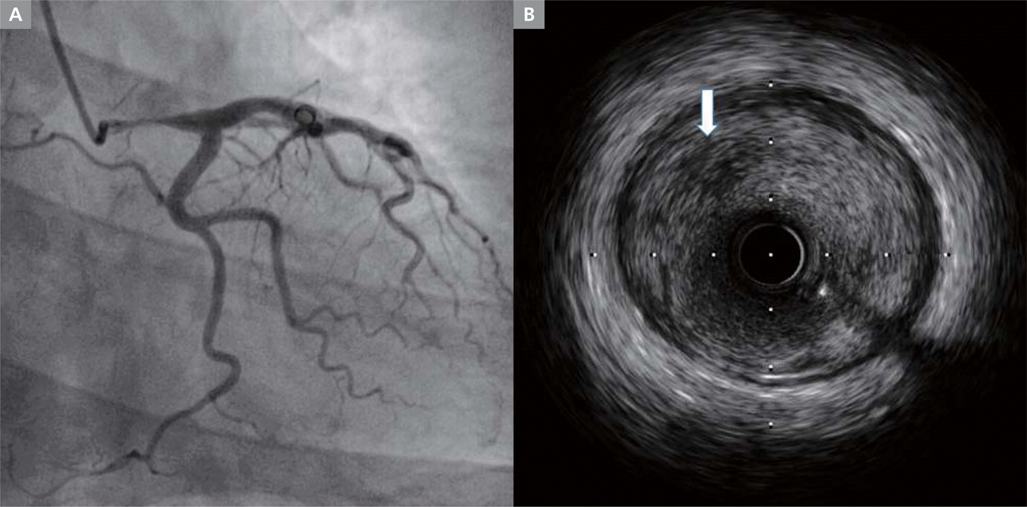
While waiting for the test results, syncope recurred in the emergency room. Routine laboratory test results were normal, except for cardiac troponin, which was slightly elevated at 0.058 ng/mL. Transthoracic echocardiography showed preserved left ventricular ejection fraction (65%) with no regional wall motion abnormality. Follow-up ECG showed type 1 Brugada pattern, which was different from the initial ECG (Figure 1A). As the time passed, follow-up ECG was changed to type 2 Brugada ECG pattern (Figure 1B). Although the cause of syncope was assessed as Brugada syndrome, coronary angiography was performed to rule out ischemic heart disease. Coronary angiogram showed significant stenosis in the left main coronary artery (LMCA; Figure 2A; Movie 1). Intravascular ultrasound (IVUS) of the LMCA showed lipid-rich necrotic cores with vulnerable plaque (Figure 2B; Movie 1). Percutaneous coronary intervention (PCI) with sirolimus-eluting Orsiro stent was performed at the LMCA. After then, syncope did not recur, and Brugada-type ECG did not appear during the 1-year follow-up period.
Figure 2.
(A) Coronary angiogram showing significant stenosis in the left main coronary artery. (B) Intravascular ultrasound (IVUS) showing lipid-rich necrotic cores (arrow) of vulnerable plaque.

Syncope is common and has many possible causes. Previous studies showed that cardiac diseases account for 8.3%–23% among all causes of syncope.1–3 Syncope is relatively common in Brugada syndrome with coved- or saddle-back type ST-segment elevation in the right precordial leads (28%).4 The guideline recommends the use of implantable cardioverter defibrillator (ICD) when the cause of syncope is unknown even after sufficient examination in patients with type 1 Brugada syndrome with syncope.5 In our case, although the patient was asymptomatic for ischemic heart disease, LMCA stenosis was found on examination while evaluating the cause for syncope.
In patients with ischemic heart disease, syncope as initial presentation is very rare.6 Syncope is usually caused by cerebral hypoperfusion.7 In situations where hemodynamic changes may occur, sudden collapse of the coronary artery blood flow may lead to syncope before chest pain has occurred. Therefore, the evaluation for ischemic heart disease in younger patients without typical symptoms, such as chest pain, may be ignored, even if they have a relatively clear cause of syncope.
We speculate that the cause of syncope is likely to be LMCA disease because syncope did not occur after treatment with PCI at LMCA stenosis, and Brugada-type ECG has also not been observed during the 1-year serial follow-up period.
The clinical implication of Brugada-type ECG in this patient is unclear. LMCA hypoperfusion might have caused the Brugada-type ECG change. Alternatively, it might be just a bystander in patient with LMCA, but Brugada syndrome may develop over time. Therefore, long-term follow-up is necessary to determine the clinical significance of Brugada-type ECG in this patient.
This is the first case of LMCA presenting as syncope with Brugada-type ECG in a young patient, and it reminded us to consider ischemic heart disease as the cause of syncope with Brugada type ECG.


